AI shows its value in mammography screening
A study has shown that artificial intelligence can be safely used to support mammography to spot breast cancers and may spot more tumours than the current standard approach.
The Swedish trial published in The Lancet Oncology found that one expert reader using ScreenPoint's Transpara AI to look at mammograms was at least as good, and possibly better, than the current standard of two expert readers.
It is one of the most comprehensive assessments of AI in this setting to date, involving more than 80,000 women attending screening clinics in Sweden who were randomised to either the AI or the control group.
Detection rates were six per 1,000 women screened with the AI, compared to five per 1,000 in the control group, with 244 and 203 cases found, respectively. The false-positive rate was 1.5% in both the AI group and the group assessed by two experts.
While the authors of the study concluded that the results are not enough on their own to confirm that AI is ready for routine use in mammography screening, the large, prospective study adds to a growing body of retrospective analyses that suggest the technology has a role to play.
Use of the AI also resulted in a significant reduction in the workload faced by the screening centres, almost halving the screen time needed, which could be an important finding for healthcare systems like the UK NHS that are struggling with workforce issues.
The NHS is facing a 30% shortfall in clinical radiologists, leading to longer waiting times and worse health outcomes, according to recent figures from the Royal College of Radiologists.
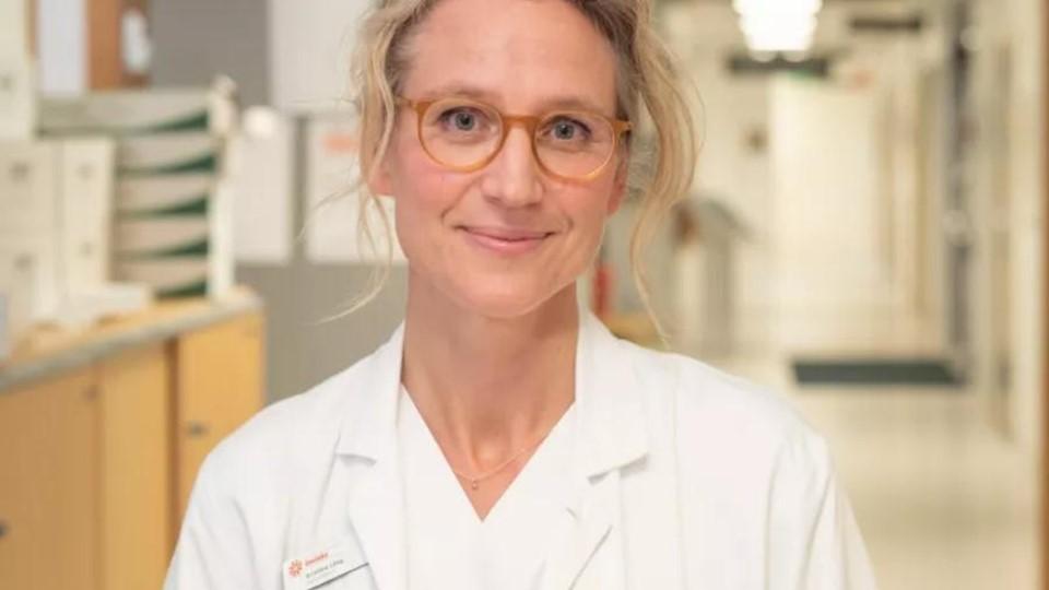
"The greatest potential of AI right now is that it could allow radiologists to be less burdened by the excessive amount of reading," said lead author Dr Kristina Lång from Lund University in Sweden.
"We need to see whether these promising results hold up under other conditions, for example, with other radiologists or other AI algorithms," she added.
The positive result means that the study will continue to accrue around 100,000 patients, with all subjects followed for at least two years to see if there are any differences between the two groups in the rate of interval cancer – breast cancer found during the three years after a normal result and before the next screening appointment – per 1,000 screens.
Prof Stephen Duffy of Queen Mary University of London (QMUL), an expert in cancer screening who was not involved in the trial, said it was high-quality work that could be of considerable importance to screening programmes.
He cautioned that technology-driven increases in detection might result in over-detection of relatively harmless lesions, but said the authors will look specifically at this issue in further follow-up.
"The results of this paper include an increase in detection of ductal carcinoma in situ, which is thought to be potentially over-diagnosed," said Prof Duffy. "It would have been helpful to see the distribution of cancers detected by grade and biological subtype. If these were similar between the artificial intelligence group and the control group, fears of overdiagnosis would be lessened."
In the UK, the NHS is currently trialling whether AI should form a part of routine mammography services.
Earlier this year, the prospective LIBRA study started at Leeds Teaching Hospitals NHS Trust, deploying Kheiron Medical's Mia AI alongside two human readers. The aim is to see if it will be possible to replace one of the human readers in future.


8c10.jpg)